![[Magnetometer]](magnetometer.gif)
Figure 10: Basic SQUID magnetometer
[Previous] [Table of Contents] [Next]
A SQUID is capable of detecting a change in magnetic field without any extra equipment. However such an arrangement is hardly ever used, for two main reasons. Firstly, any bulk superconducting material present within the SQUID would effectively screen out the field been measured. Secondly, to block out any external fields the SQUID must be operated within a superconducting shield.
In practice measurements are obtained by linking the SQUID to a flux transformer. Such a circuit consists of some type of pick-up coil linked to a secondary coil, which in turn is closely connected to the SQUID loop. Other quantities can be measured by replacing the flux transformer with other types of transducer.
A magnetometer measures the magnitude of an applied magnetic field. In this case, the flux transformer is a simple two-coil DC transformer (Figure 10). Normally both the pick-up and secondary coils are superconducting and therefore need to be cooled. The pick-up coil is placed in the field to be measured, causing a field to be set up by the secondary coil, which in turn is detected by the SQUID.
Figure 10: Basic SQUID magnetometer
A field change of ![]() B will result in a current
change of
B will result in a current
change of ![]() i, in the circuit
i, in the circuit
| (20) |
| (21) |
where n is the number of turns in the pick-up coil. If we model the pick-up coil as a long solenoid then the following equation holds
| (22) |
where l is the length of the solenoid, ![]() the
permeability of space inside the solenoid and
the
permeability of space inside the solenoid and ![]() 0 the
permeability constant. Using these equations to differentiate
0 the
permeability constant. Using these equations to differentiate ![]()
![]() with
respect to A yields the result that for maximum energy flux transfer
L1 = L2. This causes a problem. It is desirable
to have the pick-up coil as large as possible to get the best detection.
However, we also would like L1 = L2. As we
increase the size of the secondary coil it becomes increasingly difficult to
link it to the SQUID. Multiple-loop SQUIDs address this problem.
with
respect to A yields the result that for maximum energy flux transfer
L1 = L2. This causes a problem. It is desirable
to have the pick-up coil as large as possible to get the best detection.
However, we also would like L1 = L2. As we
increase the size of the secondary coil it becomes increasingly difficult to
link it to the SQUID. Multiple-loop SQUIDs address this problem.
A gradiometer arrangement is essentially the same as a magnetometer except that the pick-up coil is in fact two or more coils. Consider the arrangement shown in figure 11. The two pick-up coils are wound in opposite senses. If a uniform field is applied the flux changes in the two coils will be equal and opposite, giving a net flux change of zero. A magnetic field that is of different magnitude at the two sites of the coils will cause a non-zero total flux change, changing the current flowing in the circuit. This device is therefore a measure of the field gradient in the direction to the two pick-up coils.
![[First-order gradiometer]](gradiometer1.gif)
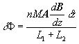
Figure 11: First order diagonal gradiometer measuring
dBz/dz tensor component
![[First-order
off-diagonal gradiometer]](gradiometer1-off.gif)
Figure 12: first-order off-diagonal gradiometer, measuring the
dBx/dz tensor component.
![[Second-order gradiometer]](gradiometer2.gif)
Figure 13: second-order diagonal gradiometer.
The gradient of a vector field is a 3x3 tensor. The arrangement shown in figure 11 is suitable for measuring the diagonal components of this tensor. To measure the off-diagonal components arrangements such as that shown in figure 312 must be used.
In figure 11 the coils are aligned in the z direction
separated by a small distance ![]() z, enabling measurement of the
dBz/dz component of the gradient. Adapting equation 2 we can
write the flux change at the SQUID as follows
z, enabling measurement of the
dBz/dz component of the gradient. Adapting equation 2 we can
write the flux change at the SQUID as follows
 |
(23) |
where A is now a vector representing the area of the coil.
It is also possible to measure higher order gradients of the magnetic field. Consider two sets of primary coils, as shown in figure 11, connected back to back. This will enable d2B/dz2 to be measured. Such a circuit is shown in figure 13; the two middle coils have been combined into a single one. Although in theory it is possible to build a circuit to respond to any order of gradient, in practice only those measuring up to the 3rd order have been realised.
The basic line of thinking behind an ammeter or voltmeter using SQUID technology is to insert a coil directly into the superconducting loop. This can then be used to control a change in the magnetic flux of the ring. The general case of this concept is to apply the coil with the external circuit shown in figure 14.
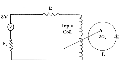
Figure 14: Generalised circuit diagram for electrical measurement using a
SQUID.
These devices pioneered by Harvey (1972) are widely accepted to be more than 200 times superior than conventional DC comparators. A wire is placed through a superconducting metal tube and the DC current through it is allowed to set up a screening supercurrent that runs in one direction on the inside of the tube and the opposite direction on the outside of the tube. The ingenious part of this design is that the spread of the screening current on the outer tube is independent of the position of the wire in the tube. We now insert another wire into the apparatus (the test wire) and we can measure the B-field created by the screening current on the outside of the tube. We can now vary the currents until the B-field is zero, as detected by the SQUID. This technique has been used to establish a 1:1 current ratio with an accuracy of 1 part in 1012. Later designs saw the tube being bent around onto itself (toroidal), but the ends do not make electrical contact. These methods are effective but it must be noted that any external field must be very stable and the flux transformer extremely rigidly mounted Superconductor shielding is a priority for high resolution. This work has lead to a portable secondary voltage standard (Gallop 1977) using a small 1 litre liquid helium cryostat.
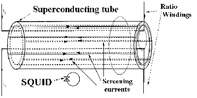
Figure 15: Pictorial representation of DC current comparator.
The high sensitivity and available resolution of SQUID circuits make them an ideal calibration tool.
Kamper and Simmonds (1973) used their microwave SQUID to calibrate a RF attenuator. Radiation from a klystron was passed through isolators, a directional coupler and finally a waveguide. The critical current through the junction was exceeded by adjusting the microwave power (of order picowatts) in small increments. The reflected microwave power was found experimentally to vary sinusoidally with the current. Hence if the detector responds to the average reflected power we find the mean detected power is equal to a constant plus a Bessel function. The attenuator is connected between the SQUID and the stable RF source. The number of zero crossings in the detected power can be used to measure changes in settings on the attenuator. The pioneers calibrated the RF attenuator using a table of Bessel functions.
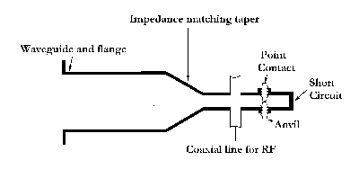
Figure 16: Experimental set-up of an AC attenuator
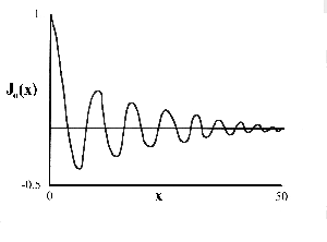
Figure 17: General Bessel function.
The high sensitivity of SQUIDs enables measurements of very weak magnetic properties on small samples to be made. When these methods were first employed in the 1970�s, much work was to follow on the superfluidity of 3He-A and 3He-B allowing detailed analysis of the susceptibility and transverse nuclear magnetic resonance (NMR). An idea of the precision of data available is given in the graph (figure 18).
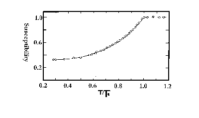
Figure 18: The susceptibility of 3He at low temperatures.
The basic design of a SQUID magnetometer measuring
susceptibility is to wind the pick-up coil of a flux transformer around the
sample. The susceptibility can then be written in terms of the flux induced in
the coil. With an applied filed of 0.1T it should be possible to detect a
susceptibility of ![]() m
m ![]() 10-12. In reality to
secure this accuracy ambient field fluctuations must be reduced by a factor of
105. This is achieved by a second pick-up coil forming an �astatic�
pair of separated counter wound coils. The whole apparatus is then placed in a
long cylindrical superconducting shield. Figure 19 shows a pictorial
representation of the typical apparatus.
10-12. In reality to
secure this accuracy ambient field fluctuations must be reduced by a factor of
105. This is achieved by a second pick-up coil forming an �astatic�
pair of separated counter wound coils. The whole apparatus is then placed in a
long cylindrical superconducting shield. Figure 19 shows a pictorial
representation of the typical apparatus.
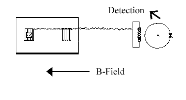
Figure 19: Experimental set-up of apparatus.
Kamper and Zimmerman (1971) pioneered experiments in this field. They thought of the SQUID as a parametric amplifier whose noise temperature is lower than its operating temperature. These devices represent new standards of precision in the range 0.001-4.2K.
A typical circuit for noise voltage thermometry is shown
(figure 20). A thermometric element (resistance R) is incorporated into a
superconducting RF biased magnetometer. The bias voltage due to the current
io causes the quantum phase difference across the weak link
(![]() (t)) to precess at the Josephson frequency;
(t)) to precess at the Josephson frequency;
![]()
The result is an AC output voltage at V0 from
the detection circuitry. The time-dependent Johnson noise appears in series
with R and is added to V0. This noise frequency modulates the
carrier signal V0 to a degree dependent on the instantaneous value
Vn(t) of the noise voltage. Nyquist�s theorem is used and via an
application of FM theory (Burgess, 1967) the linewidth
![]() V can be written
in terms of the absolute temperature of the device. The accuracy of measurements
achieved so far is in the region of +/- 1mK at 20 mK. It is difficult to
predict the minimum temperature measurable.
V can be written
in terms of the absolute temperature of the device. The accuracy of measurements
achieved so far is in the region of +/- 1mK at 20 mK. It is difficult to
predict the minimum temperature measurable.
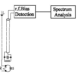
Figure 20: Experimental set-up of a Johnson noise thermometer
The scanning SQUID microscope is an extremely sensitive instrument for imaging local magnetic fields. It scans the SQUID relative to the sample to image the local magnetic fields with unprecedented sensitivity. As with most scanning probe microscopes the probe is moved about using piezoelectric scanning elements. However a scan area of a few hundred microns square is very difficult to achieve with these piezoelectric crystals operating in a cryogenic environment. Another design problem is that the SQUID has to be very close to the sample surface to obtain optimal resolution.
The design (not shown) by Vu et al provides the highest possible resolution that�s necessary for imaging the magnetic field vortices present outside a super-conductor. This design has a large reservoir of liquid helium in the middle and both SQUID and sample are cooled to liquid helium temperature. The SQUID is directly linked to a stepper motor, which allows the SQUID to be scanned across the sample. In this example the SQUID is placed in series with an inducting loop (50pH). It is made from pure Niobium (Tc=9.1K); its critical current is 17uA. Both this design and Magnetic Force Microscopy [14] are complementary methods for probing the magnetic structure of various materials. These two designs both have the disadvantage of significant interaction between the sample and the probe thereby changing the studied sample. The best design of SSM so far has to be the one by Kirtley et al [15].
This design is an improvement on the aforementioned because
the SQUID is mounted on a brass cantilever [16], which
allows some vertical movement of the SQUID. The SQUID can now be run in direct
contact with the sample with the cantilever mechanism absorbing some of the
unevenness and this proximity to the sample improves the resolution
considerably. The whole of the above mechanism is contained in a thin-walled
stainless steel tube, which is vacuum-sealed and connected to imaging
electronics. The whole of this tube is contained in a
![]() -metal-shielded Dewar (i.e. shielded to magnetic
fields) full of liquid helium and suspended from the ceiling by chords to
eliminate any vibrations. The SQUID itself is a thin-film DC SQUID with two
Josephson links separated by a ceramic layer, a picture of which can be seen
in figure 21, [17].
-metal-shielded Dewar (i.e. shielded to magnetic
fields) full of liquid helium and suspended from the ceiling by chords to
eliminate any vibrations. The SQUID itself is a thin-film DC SQUID with two
Josephson links separated by a ceramic layer, a picture of which can be seen
in figure 21, [17].
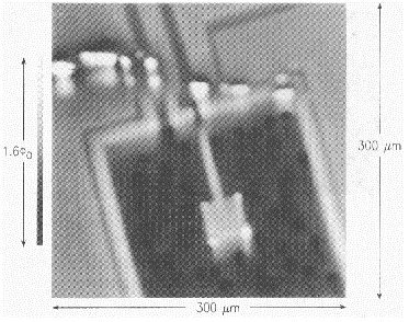
Figure 21: SSM image of a SQUID
This image of a high-Tc thin-film SQUID was produced with the SSM described above. The outlines of the SQUID are visible because the supercurrents circulating the device produce a characteristic magnetic field, which screens out the background field. The SSM being discussed use this device in conjunction with a 10um pick-up loop, very small components such as these are made by electron-beam lithography techniques [18].
Until recently the role of SQUIDs has been limited in making many geophysical measurements. Today, however the SQUID has many uses, not least the measurement of the thickness of the Earth�s crust by considering how it responds to incident low-frequency electromagnetic disturbances propagating from the ionosphere. To gain useful measurements of this kind the levels of magnetic noise in a quiet open site have to be considered. In a remote site the only noise that has to be considered are the intrinsic fluctuations of the Earth�s magnetic field. These fluctuations are frequency dependent and occur between 0.1-10Hz with an approximate magnitude of 10-11 THz-1 at 1Hz. This noise level is about three orders of magnitude less than present in a normal laboratory and should be low enough to allow the SQUID to operate reasonably well. To measure the thickness of the earth�s crust the electromagnetic disturbances discussed above are treated as plane waves (to a good approximation). By measuring both the component of the magnetic field parallel to the surface and the resistivity of the surface the impedance of the earth�s surface can be determined. From this the crust thickness can be determined by the following formula [19]
![]()
where f is the frequency in Hz and 1/![]() is the
resistivity of the earth (ranges between 1 Ohm-m and 104 Ohm-m).
The fact that much better data has been collected using a superconducting
magnetometer in a portable cryostat than with the traditional search coil or
optical pumping magnetometers has led to much more widespread use in oil
prospecting and earthquake prediction.
is the
resistivity of the earth (ranges between 1 Ohm-m and 104 Ohm-m).
The fact that much better data has been collected using a superconducting
magnetometer in a portable cryostat than with the traditional search coil or
optical pumping magnetometers has led to much more widespread use in oil
prospecting and earthquake prediction.
The measurement of biologically produced magnetic fields was unknown of before the invention of SQUIDs. This is because the fields in question are of such a small magnitude, typically 1nT to less than 1pT, that no other detector is sensitive enough. There are many magnetic fields which have been measured, ranging from the susceptibility of tissue to applied magnetic fields to ionic healing currents and those associated with neural or muscle activity.
With fields of such a small magnitude, ambient magnetic noise is of comparable magnitude to the signal being measured. Therefore using a SQUID magnetometer in an unshielded environment is not possible and other techniques need to be employed. Figure 2 shows relative noise levels.
By far the largest area of study within biomagnetisum is brain imaging. Most existing non-invasive brain imaging methods such as Computerised Tomography (CT), Magnetic Resonance Imaging (MRI) and Positron Emission Tomography (PET), measure the distribution of some kind of matter and are therefore primarily a measure of structure.
Magnetoencephalography (MEG) utilises SQUIDs to measure the magnetic fields produced in the brain by ionic current flow arising from neural activity. Current MEG techniques have spatial resolutions of 1-5mm but the time resolution is between 1-5ms allowing real time imaging. This real time imaging allows research into epileptic seizures and other psychological disorders.
Figure 22 below shows the spatial and time resolution for MEG, MRI and PET imaging techniques.
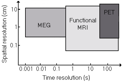
Figure 22: Comparison of the spatial and time resolutions of MEG, MRI and
PET
The main problem comes from the actual computing of the current density. For every magnetic field recorded there is an infinite number of current densities that would give rise to such a field. To overcome this the brain is considered to be a much simpler set of current sources than it actually is. A solution is then computed fitting certain boundary conditions (such as distribution having the least energy). The brain is also too complex a conductor to be considered exactly and so is approximated to something such as a homogeneously conducting sphere. Surprisingly with such harsh simplifications, an accurate current distribution can be obtained. Another method involves considering the brain to be a small number of current dipoles. Accurate 3D images of the current density within the working brain can now be readily produced.
As explained earlier ambient noise will dominate any MEG data unless precautions are taken. The most obvious solution is to enclose the whole measuring system within a magnetically screened room. This is very effective, but expensive as well as limiting and cumbersome. Often gradiometers of varying order are used to detect the field. With a gradiometer, noise will not be detected if it of constant magnitude at both of the pick-up coils. If it is assumed that the source of the magnetic noise is much further away than the signal source, this is approximately true. Therefore, with a gradiometer the signal magnitude is much stronger than the noise. Generally, the higher order the gradiometer the less noise. But higher order gradiometers are also less effective at picking up the signal. Some current systems employ computer processing to calculate a virtual second or third gradiometer output based on data from first order gradiometers. This is less effective than using real higher order gradiometers, but in many cases more practical. In some cases measurements have been made in an unshielded environment [20], although usually, too much noise is still present. Other systems use different methods to help deal with noise, such as attempting to measure it separately from the signal.
[Previous] [Table of Contents] [Next]
This document was saved as HTML from a Word 97 document and then labouriously converted from its nasty output. This document was last updated on Wednesday 28th October 1998.